 中文版
中文版 English
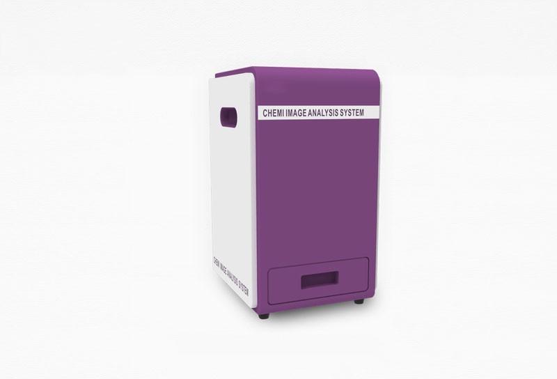
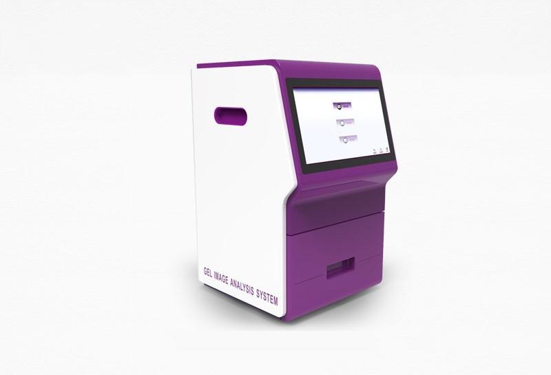
Gel imaging analysis system
manufacturer:Shandong Horde Electronic Technology Co., Ltd.
Introduction:The manufacturer of Gel imaging analysis system「Horde Electronic」 has mastered the research and development technology of Gel imaging analysis system, with more than 10 years of experience in manufacturing Gel imaging analysis system. The quality is reliable, the detection accuracy is high, and the price competitive advantage of the source manufacturer is obvious. Welcome to call for more information.
Update time:2026-06-11
- Contact: Contact Information
- Phone:008619053605658
Product Recommendation

Product Details
1. Product Introduction:
Gel imaging analysis system (UV) is suitable for the collection and analysis of images such as DNA/RNA electrophoresis gel, protein electrophoresis gel, and spot hybridization. It uses a high-resolution camera with a high-sensitivity lens to ensure high-quality imaging effects.
Two configuration models are available:
● UV imaging - high-efficiency stable ballast, rectifier efficiency η:> 80%, extending the life of the lamp, and the average number of switches is up to 40,000 times.
2. Application range:
1. Nucleic acid detection: various fluorescent dyes, such as Ethidium bromide, SYBR Gold, SYBR Green, GelSafe, GelRed, GelGreen, SYBR Safe, GelStar, Fluorescein, Texas Red, Fluorescein, Oligreen, Picogreen, GelStar labeled DNA/RNA detection.
2. Protein detection: Coomassie brilliant blue gel, silver stain gel, and various dyes Coomassie Blue, Copper stain, Zincstain, Flamingo, Oriole, Silver stain, Coomassie Fluor Orange, SYPROuby, Krypton labeled gel/membrane/chip, etc.
3. Other applications: culture dish colony counting, ELISA plate, dot hybridization, protein chip.
3. Product features:
1. High-resolution camera and high-sensitivity imported lens to ensure high-quality imaging effect.
2. Special gel cutting protective cover for large-size observation field, more comfortable and safe operation.
3. Intelligent operation software, zoom, focus, light source switch, shooting and other full-range operations through the computer, ensuring the interface is simple and intuitive under the premise of diversified functions, and easy to master imaging operations.
4. Technical parameters:
Camera: imported high-resolution scientific research camera
Camera chip: imported CCD
Effective pixels: 2592×1944 (5.03 million)
Pixel density: 16bit (65536 grayscale)
Pixel size: 5.2μm×5.2μm
Photosensitivity: 0.78
Signal-to-noise ratio: 69.8dB
Read noise: 5.1e-RMS
Detection sensitivity: can detect nucleic acid as low as 0.02ng
Lens: imported high-transparency 6x auto zoom lens, F1.0, 8-48mm; one-button auto focus function, can automatically focus on the sample being photographed, no manual adjustment required
Filter: imported 590nm, other filters optional
Transmitted UV: standard 302nm, using high-efficiency rectifier, rectifier efficiency η: >80%, 1.13 lamp, average power on/off times up to 40,000 times; (optional 254nm, 365nm)
Transmitted white light: standard three-color temperature LED uniform light panel
Transmitted blue Light: optional 470m
Falling UV: optional 254nm/365nm/470nm
Falling white light: high-brightness LED cold light source on both sides
UV transmission area: 210×260mm, visible light transmission area: 210×260mm
Glue cutting device: standard dedicated glue cutting device
Module control: highly programmed, can realize lens module (zoom/focal length/aperture), transmission light source and reflection light source, glue cutting light source full automatic modular control
Timed light off: 1-60 minutes without operation automatically turns off the UV light source, effectively extending the service life of the UV lamp
Structure: airtight without light leakage, microprocessor controlled dark box
Protection: automatic protection system when opening the door to prevent UV damage
5. Intelligent software:
1. Image capture: light source/lens/camera control, automatic/manual exposure, automatic/manual focus, image capture, image management;
2. There is a function hidden setting, and some functions can freely choose the operation key to display or not display to ensure the simplicity of the software operation interface.
3. Image processing: adjust brightness, contrast, Gamma, image rotation, image inversion, image cropping, image scaling, image staining, image merging;
4. Analysis function: automatic/manual identification of lanes, automatic/manual identification of bands, manual addition and deletion of lanes and bands, customizable molecular weight standards, and calculation of molecular weight and concentration;
5. The operating software comes with a direct printing window. Grid line function, after selection, the image interface will have evenly arranged green grid lines, and a movable purple horizontal line is specially set, and the fixed green frame line is matched with the movable purple line, which is convenient for reference and comparison of Markers;
6. There is a default image saving path, no need to save manually; you can also customize the saving path before shooting, and the picture taken this time will be automatically saved in the selected path. After shooting and exiting the software, the default path will be automatically restored next time to prevent storage confusion;
7. Other analysis functions: colony, spot hybridization clone counting;
8. Data export: The results can be exported to Excel;
9. The software is free for lifetime on-site installation, upgrade, and training, and on-site maintenance is carried out according to customer needs;
6. Configuration list
1. Imaging host, 1 unit;
2. Data cable, 2;
3. Power cord, 1;
4. Certificate of conformity/warranty card, 1 copy;
5. Transmission white light board, 1 copy;
6. Cutting cover, 1;
7. Software USB flash drive, 1;
Product's Website:http://en.huoerd.com/hxfgcxxt/1789.html


 Home
Home Phone
Phone Product
 Contact